Research
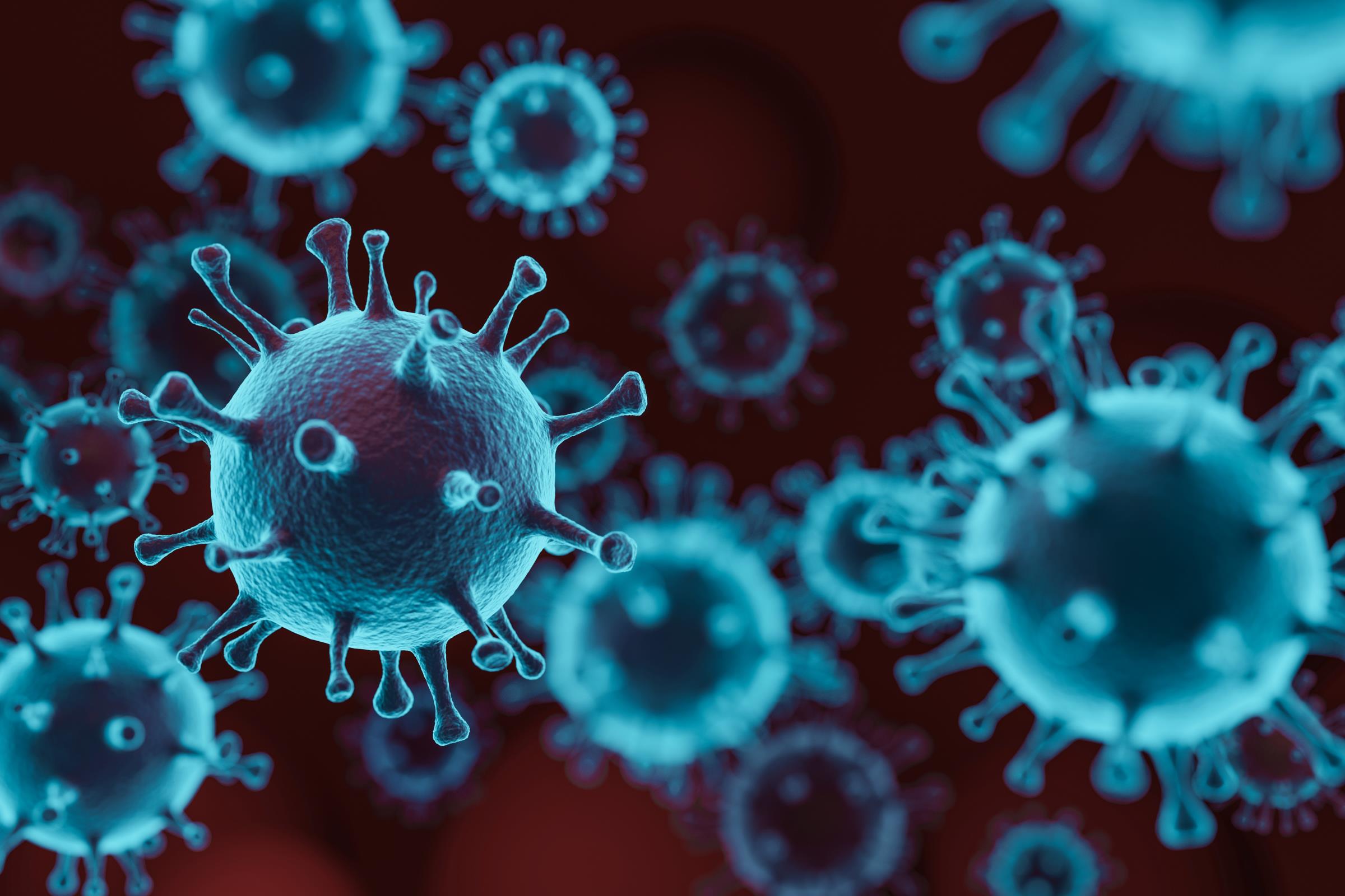
The Lindenbach laboratory is focused on the molecular biology of positive-strand RNA viruses. Key features of these viruses are:
- Many important human, animal, and plant pathogens are positive-strand RNA viruses, including coronaviruses (e.g., SARS-CoV-2), flaviviruses (e.g., dengue, yellow fever, and Zika viruses), hepaciviruses (e.g., hepatitis C virus), alphaviruses (e.g. chikungunya virus), noroviruses (e.g., Norwalk virus), and many others.
- Positive-strand RNA viruses have small RNA genomes that are directly translated as messenger RNAs (mRNAs). Because viral proteins are produced by translation of the viral genome, positive-strand RNA virus genomes are directly infectious and can be used to reboot the entire virus life cycle upon transfection into a suitable host cell. Thus, a key tool for dissecting positive-strand RNA virus replication is a functional cDNA clone, which can be used to modify the viral genome, transcribe synthetic RNAs, and recover infectious virus.
- Positive-strand RNA viruses replicate in the cytosol of infected cells, in association with “replication organelles” built by these viruses out of cellular membranes and viral and cellular proteins. RNA replication occurs via production of a complementary negative-strand RNA, which serves as a template for production of more positive-strand RNA genomes. Thus, these viruses replicate via unique mechanisms without DNA intermediates.
- RNA replication is error prone. As a result, positive-strand RNA viruses can give rise to vast genetic diversity and are capable of rapid evolution. This error-prone replication explains the incredible diversity between positive-strand RNA viruses, as well as their small genome size (≤30-kb), since replication fidelity limits the size of a genome that can be faithfully maintained.
- The genomes of positive-strand RNA viruses thus serve multiple roles in the virus life cycle: as mRNA, as template for RNA replication, and as the genetic material packaged within virus particles. Yet viral genomes are not passive information encoders; rather, they fold into dynamic, three-dimensional structures that regulate the viral life cycle.
Much of our work focuses on the Flaviviridae, a large and diverse family of positive-strand RNA viruses, including the flaviviruses (e.g., yellow fever virus), hepaciviruses (e.g., hepatitis C virus), pestiviruses (e.g., bovine viral diarrhea virus), and pegiviruses (e.g., human pegivirus). We ask fundamental questions about how these viruses replicate, which may lead to new antiviral strategies. We are also interested in understanding how different viruses solve the same problems in different ways, so we like to perform comparative work between viruses. Along the way, we have used alphaviruses as tools to study flavivirus and hepacivirus replication.
Leveraging Genetic Techniques to Unravel Viral Genome
Understanding how viruses use their limited repertoire of proteins to navigate shared steps of the viral life cycle is a fundamental question in virology. These steps of cellular entry, reproduction, assembly, and release rely on conserved proteins like proteases, helicase, viral envelope or membrane proteins. To understand how each of these proteins drive the viral life cycle, we use genetic tools to remove or disrupt specific genes and assess how the virus does or does not function.
Virus Assembly
We identified a number of conserved residues in the HCV NS2 protein that are important for virus particle assembly [1]. Specifically, mutations at these sites yielded viral genomes that could replicate in RNA-transfected cells but were unable to produce infectious virus particles. We then selected for revertant viruses that overcame these defects, which revealed genetic interactions between NS2 and the E1-E2 glycoprotein and NS3-4A enzyme complexes that are important for virus assembly. To follow up, we developed methods to biochemically capture NS2-containing complexes from virus-producing cells, which confirmed that NS2 forms critical interactions with the E1-E2 and NS3-4A complexes [2]. More recently, we have undertaken a similar approach to examine the role of NS3-4A in virus particle assembly and to map residues that mediate interaction with NS2.
To examine the cell biology of HCV particle assembly in greater detail, we developed methods to fluorescently label functional core protein in virus-producing cells [3]. These data showed that core protein is rapidly trafficked to the surface of lipid droplets, which appear to associate with sites of virus assembly near the interface between the endoplasmic reticulum (ER) and lipid storage droplets. After egress from lipid droplets, core protein is incorporated into virus particles that traffic through the secretory pathway. By examining core trafficking in our NS2 mutants, we showed that the interaction between NS2 and NS3-4A is essential for recruiting core from the surface of lipid droplets into virus assembly sites. Our current working model is that the interaction between NS2 and NS3-4A regulates the flow of RNA out of replication and into packaging
RNA replication
For many HCV NS proteins, biochemical activities have been characterized and several high-resolution crystal structures are available. However what we most lack is an understanding of how these pieces work together to form the active replication complex, and how host cofactors influence the steps of translation and replication. In collaboration with Dr. Anna Pyle, we are combining genetic and biochemical approaches to close this gap in our knowledge. Specifically, we are defining critical interactions between the serine protease and RNA helicase domains of NS3-4A [4], and examining how changes in NS3-4A conformation correlate with function. Furthermore, we have developed a novel trans-complementation system to dissect the assembly of functional replication complexes.
Long-term Goals & The Big Picture
We have established a track record of accomplishment for our work on HCV particle assembly, and are making great progress on HCV RNA replication and innate antiviral defense. One long-term goal is to extend our studies to other members of the Flaviviridae to identify common themes and differences within this virus family. To this end, we have initiated studies to understand the role of NS proteins of yellow fever virus and dengue virus in replication and virus assembly. A second long-term goal is to develop new reagents to study HCV. To this end, we are developing novel methods to rapidly screen and identify additional HCV isolates that replicate in cell culture.
Molecular Determinants of Hepatitis C Virus Replication and Assembly
Multiplex Genome Editing to Dissect Complex Viral Phenotypes
Essential early events in the flavivirus lifecycle