As an assistant professor in the Radiology & Biomedical Imaging Department at Yale School of Medicine, Anne Marie Boustani, MD, interprets diagnostic and molecular imaging for the management of cancer and other diseases. Boustani joined the department five years ago after completing a fellowship in nuclear medicine. But before she attended medical school and trained as a radiologist, she followed a different path, completing a two-year graduate program in medical and biological illustration at Johns Hopkins University. Now, she is using her skills as an MD and an artist to create medical images for her peers.
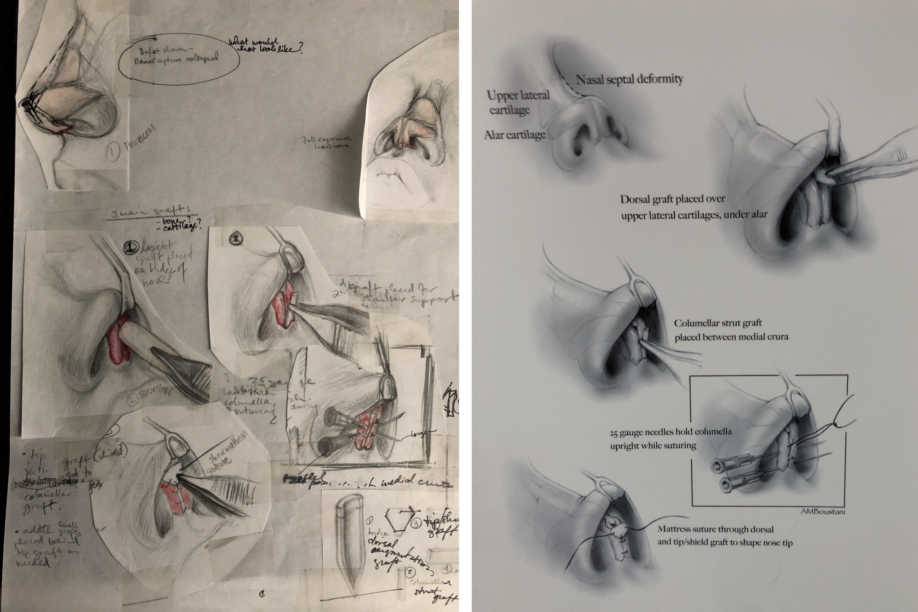
At Johns Hopkins, Boustani learned how to sketch quickly while observing surgery. “I would read about the surgery later and then refine the drawing,” she said. “I had majored in art in college. I also loved science,” Boustani said, “and while I was at Johns Hopkins I was drawn to gross anatomy and histology courses. I found myself wanting to learn more. That’s when I decided I wanted to go to medical school and become a physician.”
During medical school, Boustani put aside her artwork, except for the occasional doodle, until recently, when she began to illustrate research papers for her colleagues. “I love being a physician, but I also love that I have this other skillset that allows me to collaborate in a creative and productive manner,” she said.
Julius Chapiro, MD, PhD, co-director of the radiology department’s Interventional Oncology Research Lab, is an expert on the use of magnetic resonance imaging (MRI) in liver tumor models. While visiting the nuclear medicine reading room recently, he mentioned how difficult it was for him to explain his research in simple terms. Boustani offered to help. After reviewing several research slides, she made a draft illustration that showed the sequence of his research, beginning with the growth of liver cancer cells and ending with a drawing of a miniature MRI machine.
“She was responsive, fast, and she added a lot of value to the paper,” Chapiro said. “Our review process was surprisingly unproblematic, and we got this paper accepted in a top journal in no time. I do credit Anne Marie’s figure a lot for it.”
Boustani receives author credit in line with the value added by her illustrations. “Being an MD helps. I understand what people want and can help answer questions that arise through illustrations,” she said. “I don’t think we use pictures enough to explain complex research.”
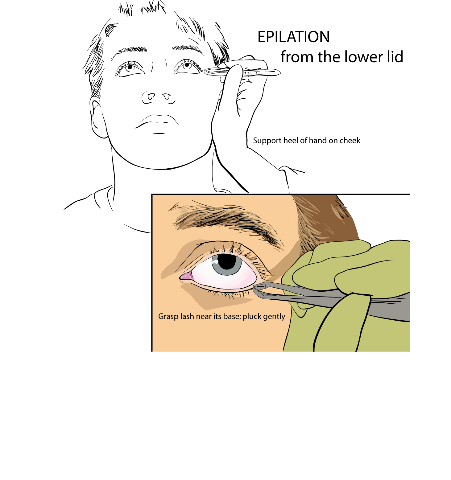
Her skills are also being used to help with patient experience. “If I want to teach patients about a procedure, how can I best do that? It’s not just about doing anatomical drawings; it’s about being able to create clear pictures that show a process,” she said.
“While all my faculty are accomplished imagers, Anne Marie has the additional rare gift of illustration,” said Chair Rob Goodman, MD. Goodman said the Department of Radiology and Biomedical Imaging is exploring ways to offer Boustani’s skills more widely as part of a medical illustration service. To find out more, please contact the department at 203-785-6938.