Research: Imaging Brain Activity
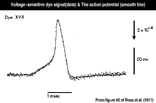
The figure illustrates the voltage-sensitive dye signal (dots) and the action potential (smooth line) measured simultaneously from a squid giant axon. The two signals follow each other precisely providing one kind of evidence that this dye signal is potential dependent.
Ross, W.N., B.M. Salzberg, L.B. Cohen, A. Grinvald, H.V. Davila, A.S. Waggoner, and C.H. Wang (1977). Changes in absorption, fluorescence, dichroism, and birefringence in stained giant axons : optical measurement of membrane potential. J Membr Biol, 33, 141-183.
One reason the brain is difficult to study is that many individual neurons or brain areas are active at once; conventional techniques allow one to monitor only one or a few neurons or locations at a time. We have worked on developing several variations of optical methods for measuring brain activity; these include organic voltage and calcium sensitive dyes and fluorescent protein voltage sensors (GEVIs). Concurrently we have applied these sensors to measuring activity in Aplysia, salamander, rat, and mice brains. At present we are concentrating on improving fluorescent protein voltage sensors and using them to measure activity in the mouse olfactory sensory system.
- A protein activity sensor has the important advantage that it can be specifically expressed in an individual cell type in the brain. For the past 15 years or so we have been working to improve protein sensors of membrane potential.
- Each pixel in a population recording receives light from a large number of neurons and processes (e.g. from an area of brain 50µm x 50µm to 200µm x 200µm) and thus each signal represents the average of a population of neurons. There are several interesting aspects of vertebrate brain function where populations are involved. One example is the organization of visual cortex into modules such as ocular dominance columns. Another is the synchrony and oscillations that accompany sensory processing. A third is maps of the input to glomeruli in the olfactory bulb where 10,000 receptor neurons with identical olfactory receptor protein converge onto a single glomerulus. For studying phenomena of this type population recordings should be useful.
- In the organic voltage sensitive, we use the dyes to follow the spike activity of individual neurons, and in favorable preparations about 500 individual neurons can be monitored simultaneously. In ganglia from sea slugs (opisthobranch molluscs, Aplysia), this number is a substantial fraction of the total number of neurons present. We hope that monitoring many neurons simultaneously will improve our understanding about how nervous systems are organized to generate behaviors.
Using 2-photon microscopy we have been studying the activity of individual juxtaglomerular neurons in the mouse olfactory bulb in response to odorants and to activation of an invdividual odorant receptor subtype.